大 鼠 睪 丸 間 質 細 胞 瘤
- 內容
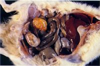
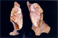
一、 病 歷 : 23 月齡 F344 雄鼠,皮毛粗剛、消瘦、無精神。 二、 肉眼病變 : 雙側睪丸腫大,剖面呈橘黃色,可見許多增生小結節,其它內臟正常。 三、 組織病變 : 睪丸實質可見許多增生之間質細胞小結節,外圍以結締組織 (圖3),這些細胞呈圓形至多角形,嗜紅性細胞質並有明顯空泡 (圖4)。 

四、 診 斷 : 大鼠睪丸間質細胞瘤 (Interstitial Cell Tumor in F344 Rats) 五、 討 論 : Fischer 344 (F344) 近交大鼠是極佳研究人類或其它動物睪丸間質細胞瘤的動物模式。因為 F344 大鼠在 18~24 月齡時, 62.5~68.4 % 發生睪丸間質細胞瘤。某些報告更高達 90 % 發生率,但其它睪丸腫瘤在 F34 大鼠少見。某些病例併發間皮瘤( mesothelioma)。
在判定究竟是間質細胞結節狀增生或良性間質細胞腫瘤時有一定標準如下,可參考:
1. WHO:肉眼睪丸可見橢圓形結節可認定為腫瘤。 2. Jubb & Kennedy:睪丸異常增生病變大於1公分直徑認可為腫瘤。 3. NTP (National Toxicology Progzam):異常間質細胞增生積聚,其直徑須大於一般細精小管之直徑始認可為腫瘤。 以上三點可作為間質細胞增生或腫瘤之鑑別。
六、 參考文獻 : 1. Davey FR. Molony WC. Postmortem observations on Fischer rats with leukemia and other disorders. Lab. Invest 23 : 327 - 334, 1970. 2. Kay S. Fu Y, Koontzww, ChenATL, Interstitial cell tumor of the testis. Amer J. Clin Path 63 :366 - 376,1975. 3. Cockrell BY, Garner FM, Animal models-interstitial cell tumor of the testis in rats. Comp Path Bull VII (2), 1976. 4. Goodman DG, Ward JM, Squire RA, ChuKC, Linhart MS, Neoplastic and nonneoplastic lesions in aging F344 rats Toxi. Appl. Pharma. 48: 237 - 248, 1979.
